Nuclear Stress Testing

Nuclear Stress Testing

A nuclear stress test is an imaging examination that evaluates blood flow through the heart muscle. It can help identify areas of the heart that may not receive enough blood during physical or medication-induced stress.

The test combines a small amount of radioactive tracer with specialized heart imaging. Results can help physicians evaluate possible coronary artery disease and determine whether additional testing may be needed.


How Does It Work?

A small amount of radioactive tracer is injected into a vein. Special imaging equipment takes pictures of blood flow through the heart muscle.

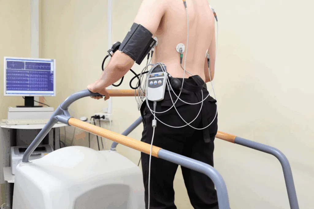
Images may be obtained at rest and again while the heart is under stress. Stress can be produced through exercise or medication when a patient cannot exercise adequately. The physician compares the images to evaluate how blood flow changes with stress.

Frequently Asked Questions

What does a nuclear stress test show?

The test shows how well blood flows through the heart muscle. It can identify areas that receive less blood during stress.The results can provide information about possible coronary artery disease. It may also help evaluate how the heart responds to treatment.

Do I have to exercise during the test?

Not always. Some patients exercise on a treadmill or another appropriate method.Patients who cannot exercise sufficiently may receive medication that increases blood flow or stresses the heart. The method is selected based on your health and ability to exercise. Your provider will explain how your particular test will be performed.

Is the radioactive tracer safe?

The test uses a carefully controlled amount of radioactive tracer. The amount and type of tracer depend on the imaging technique being used. Your medical team will consider your medical history before recommending the test. Be sure to tell your provider about pregnancy or the possibility of pregnancy.

How long does a nuclear stress test take?

The total appointment time can vary depending on the imaging protocol used. The process generally includes preparation, tracer administration, imaging, and stress testing.

Additional images may be taken after exercise or medication-induced stress. Your testing center will provide specific instructions before your appointment.

What happens if the test shows reduced blood flow?

An abnormal result does not automatically mean that a major blockage is present. Your physician will interpret the findings together with your symptoms and other tests.

Additional testing, such as cardiac catheterization, may sometimes be recommended. Your physician will explain what the results mean and what should happen next.